Applications
Longwave Ultraviolet Forensics Imaging Applications
> See Also: UV Forensics Imaging Systems: UVScanner, UVCorder and SpectraScanner
There are two main categories of UV imaging applications in forensics:
1. Seeing invisible surface features due to enhanced absorption
2. Seeing invisible features due to enhanced scattering
In other words, you get more absorption of UV radiation by trace evidence than with visible light. The evidence is easier to see with a longwave UV camera than with the unaided eye. Other types of trace evidence scatter UV rays much more than they scatter visible light. Again, you can see the evidence much easier.
Enhanced Absorption of UV
UV photons have more energy than visible (VIS) and infrared (IR) photons. Many materials and substances that reflect VIS and IR will absorb UV light. The higher photon energy makes this happen, since there is a greater chance of atomic or molecular transitions occurring. The shorter the wavelength of UV, the stronger the absorption. This property of UV makes invisible materials visible. If you have an organic material on an inorganic substrate, you can often get enhanced contrast with UV compared to regular color or infrared forensic photos. Examples of this include:
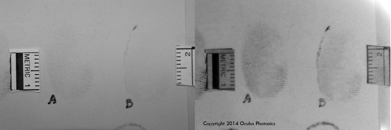
- Organic molecules found in body fluids tend to absorb UV. You can see fingerprints and bloodstains.
- Metals and inorganic materials (like cement and tile) tend to reflect UV
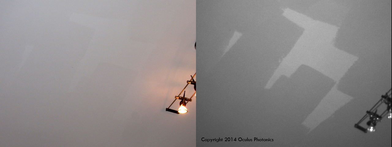
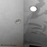
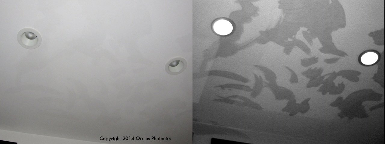
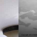
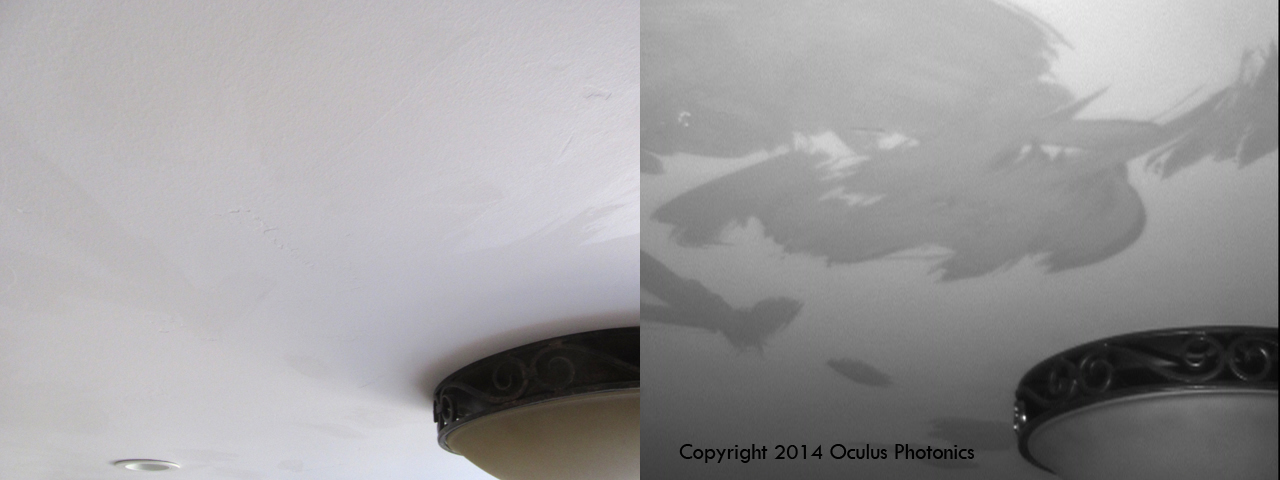
See examples of UV Absorption below:
-
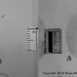
- Bloody prints
-
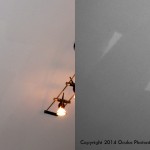
- Ceiling paint
-
- Cleaning marks
-
- Cleaning marks
-
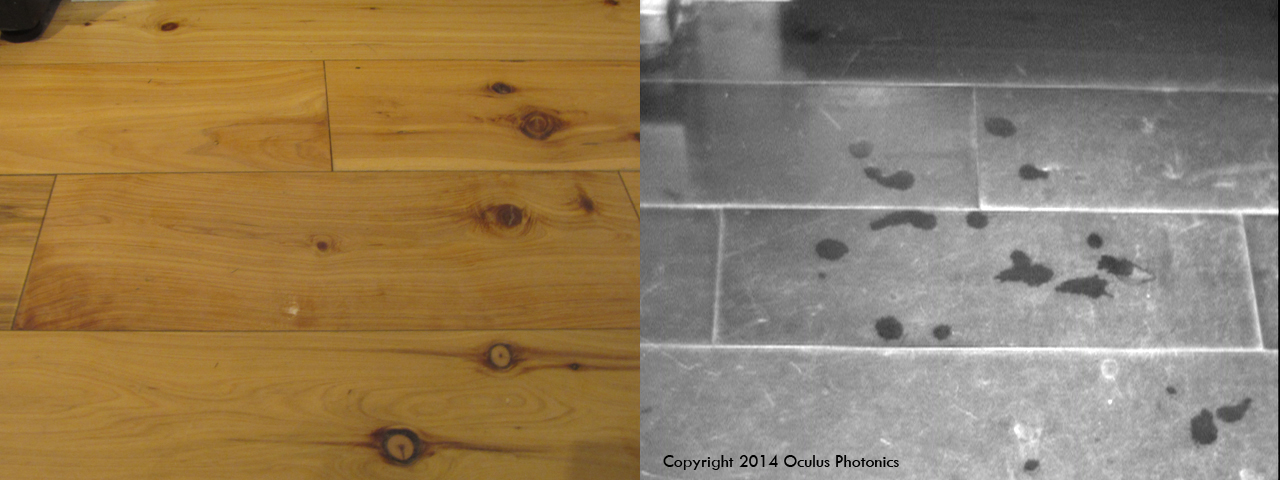
- Spit on floor
-
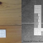
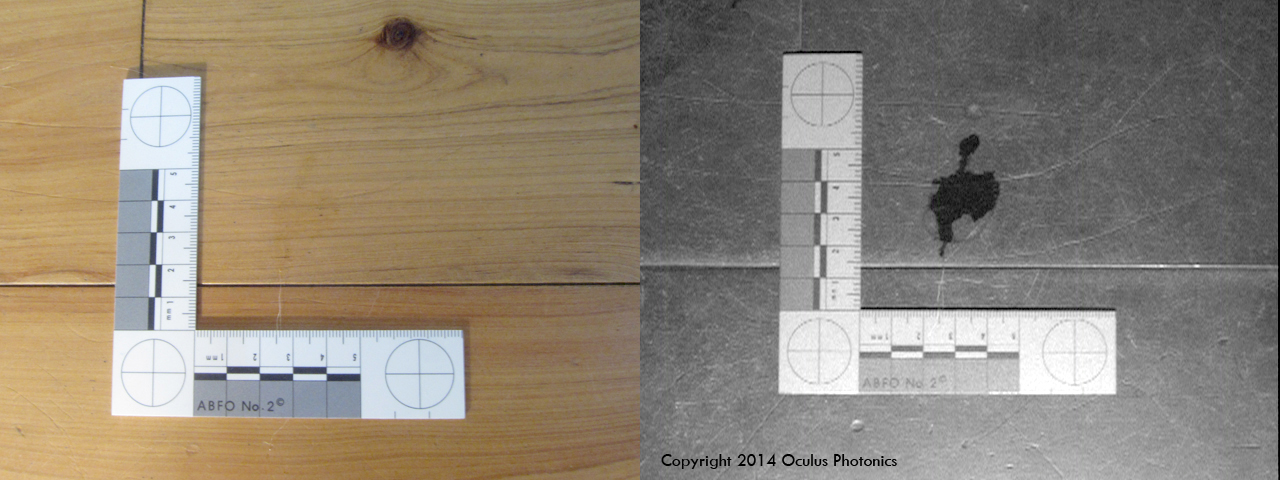
- Water & ABFO Scale
-
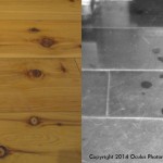
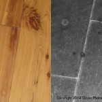
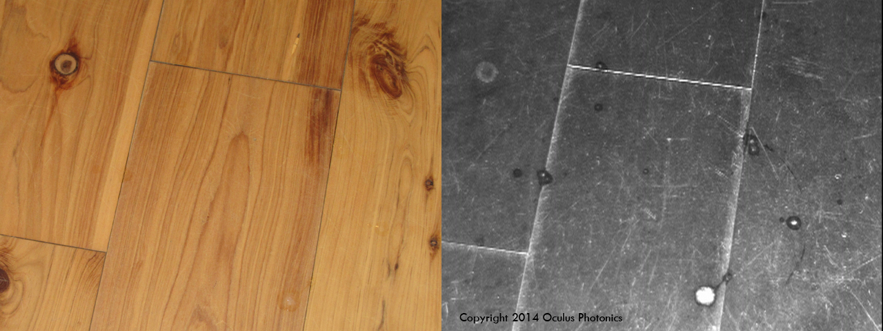
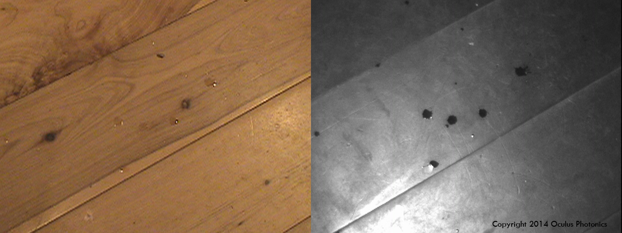
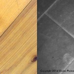
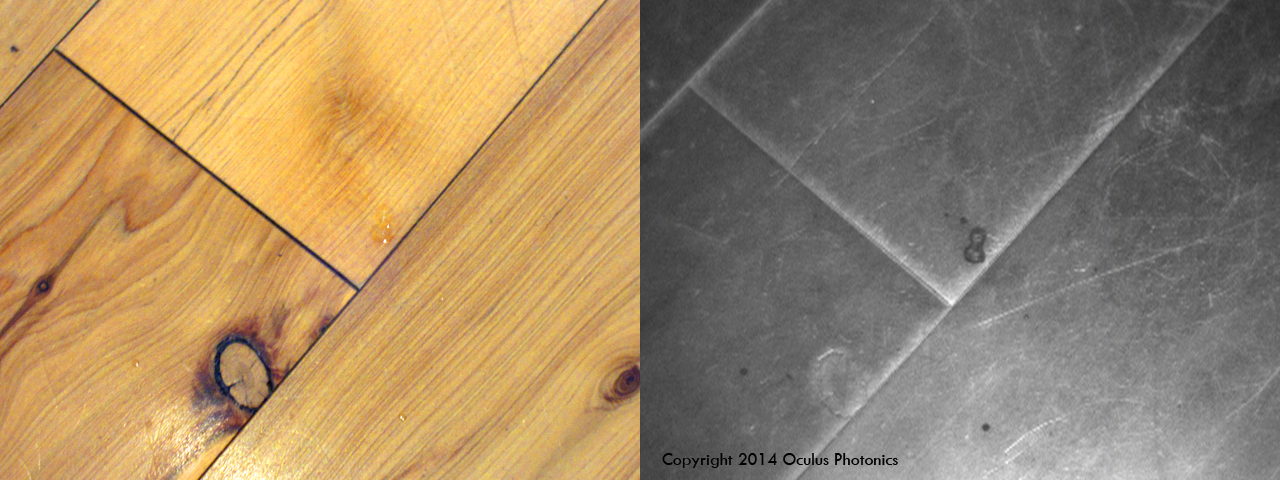
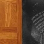
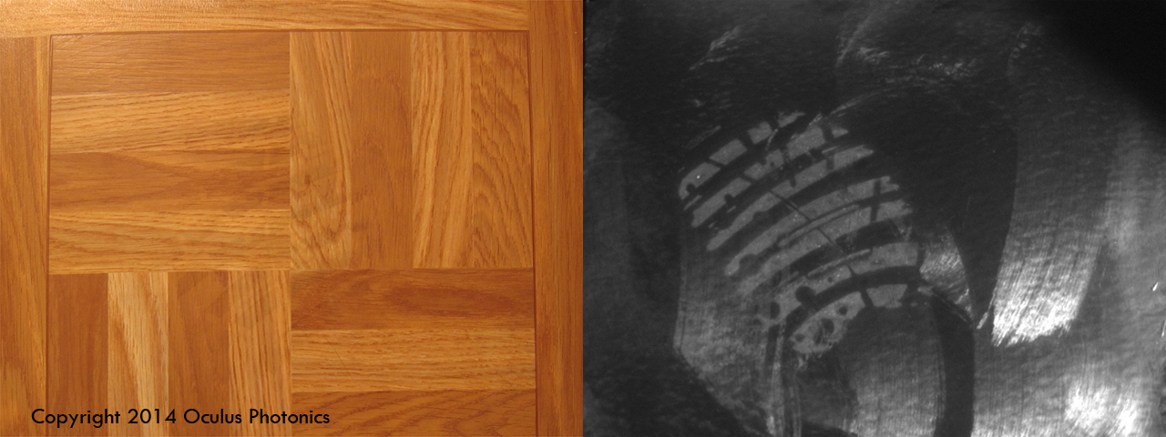
- Water on wood floor
-
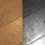
- Water on wood floor
-
- Spit on floor
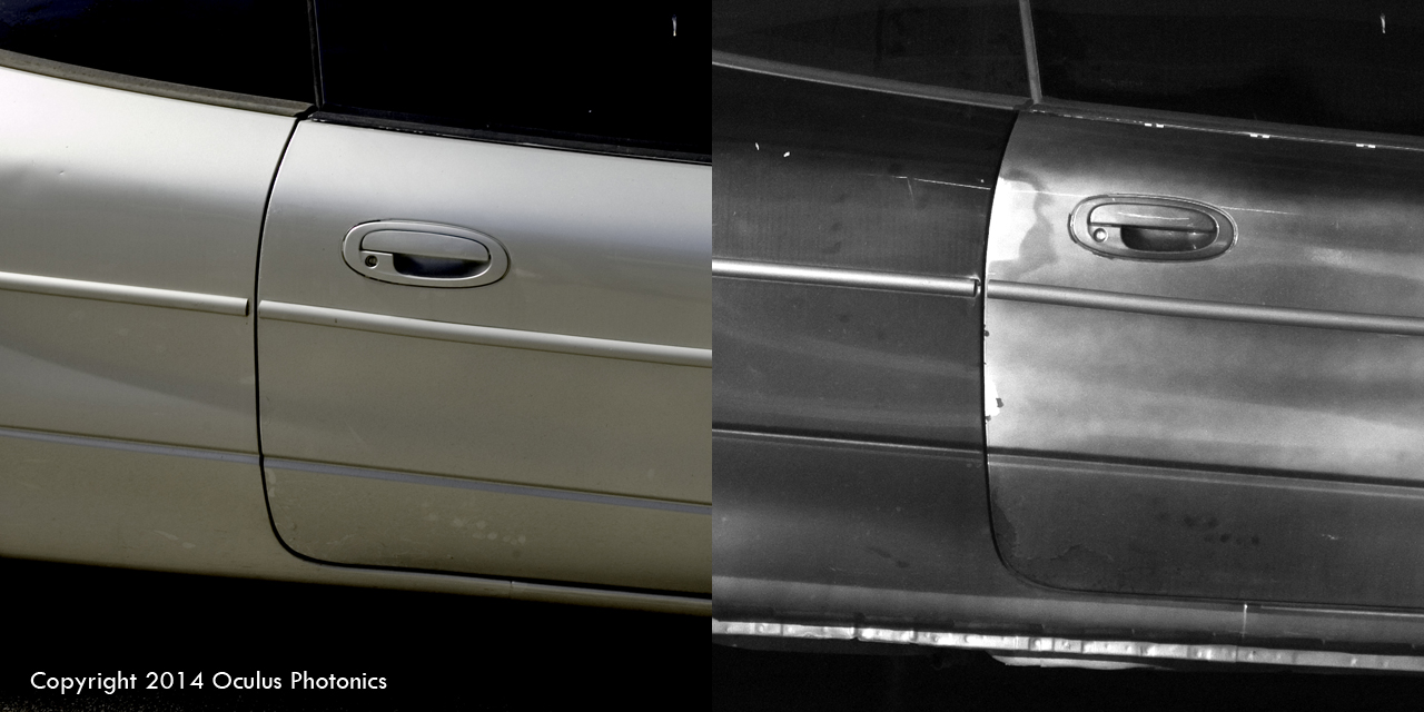
-
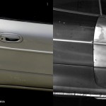
- Passenger door
Enhanced Scattering of UV
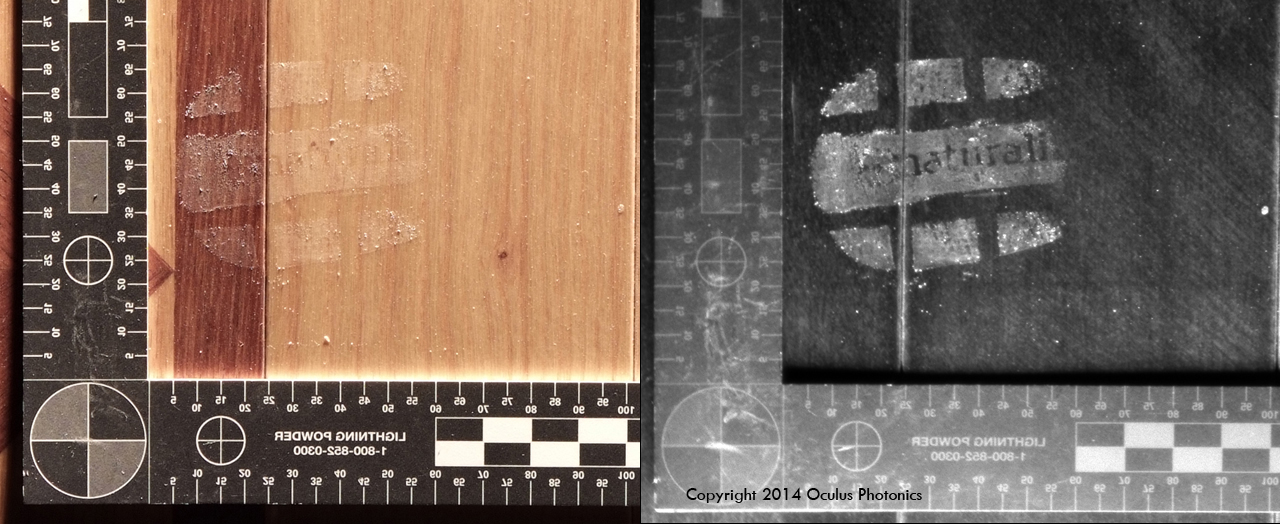
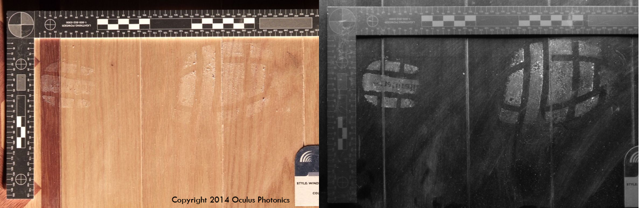
Since UV tends to be absorbed at the surface of materials, one can sometimes see surface texture that the eye will miss. This works even when the surface is translucent or transparent to VIS and IR. In addition, UV light waves have shorter wavelengths compared to visible light. UV light rays thus get scattered by surface scratches and imperfections much more readily than visible light rays.
See examples of UV Scattering below:
-
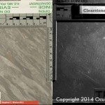
- Vinyl tile (Courtesy of Stephen Warlen)
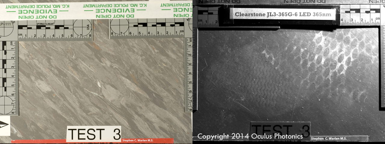
-
- Vinyl tile shoe print (Courtesy of Stephen Warlen)
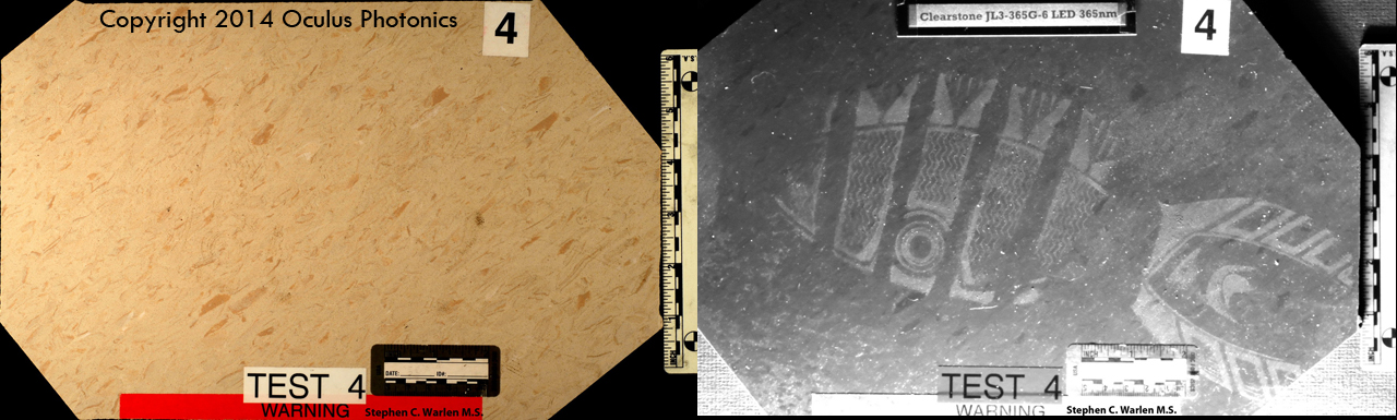
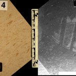
-
- Shoeprint heel (Courtesy of Rachel Leintz)
-
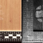
- Naturalizer shoe print (Courtesy of Rachel Leintz)
-
- Footmarks in floor wax
Notable Applications
Both the longwave UV and the shortwave UV wavebands can be used to greatly enhance the contrast in crime scene images. They can be used to quickly find evidence that is invisible both to the unaided eye and to ALS examination. Some notable applications of this include:
- Bloody fingerprints on a table (without the need for processing)
- Cleaning marks on surfaces (where blood was cleaned up)
- Dusty shoeprints on a floor (without the need for dust lifters)
- Saliva, water and other fluids staining or marking surfaces
- Repainted body panels on cars (showing repairs made after a hit-and-run accident)
Sample video taken with UVScanner
The method is non-destructive and non-invasive. Evidence can be documented without physical contact or alteration. DNA evidence can then be recovered without the problems of contamination or dilution from contact methods.
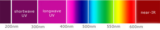
What is longwave ultraviolet radiation?
UV is a waveband, a range of “colors” of the EM spectrum. UV is invisible to human eyes – it has no “color” that human beings can describe. Images are best displayed in black and white.
For the UV waveband, one describes different “colors” of the EM spectrum by their wavelength. We use the nanometer or nm unit to describe wavelength in the UV band. The UV band can be broken into two main categories: longwave (or near) UV and shortwave UV. The longwave UV band is 300nm-400nm. The shortwave UV band is 200-300nm.
Why use it to examine a crime scene?
Longwave UV imaging sees crime scenes differently because UV light is absorbed and reflected differently than visible light. If you light up a crime scene with UV rays, and image the UV that is reflected off the scene with a UV camera, you will see things the eye can’t see. Note the distinction – this is not the same as UV fluorescence. Here UV goes in, UV comes back out and is imaged.
UV fluorescence is a “waveshifting” phenomenon. An ultraviolet excitation source is used to irradiate an area of a crime scene. Certain materials, including forensic evidence like semen, urine and fibers will glow with visible light in response to this uv excitation. The fluorescence signal is typically very weak and oftentimes requires lengthy exposures to capture photographically, which dictates the use of a tripod, which is sometimes very inconvenient to set up in tight corners.
The reflected UV methods and technologies discussed here are different. Reflected UV imaging requires that a surface be irradiated with UV radiation, which can be longwave or shortwave depending on the application. The resulting signal is quite strong and easy to record photographically. A UV-sensitive camera is then used to record a reflected uv image. Interestingly, areas that appear very bright to uv fluorescence techniques will look quite dark in reflected UV. A majority of the UV radiation is absorbed and re-radiates as visible light. The uv camera won’t see that visible light so the area looks dark.
Many crime scenes with trace evidence may exhibit little or no uv fluorescence but will reveal very sharp high contrast images of the trace evidence, either by enhanced absorption or enhanced scattering of UV. If a multiwatt longwave uv source is used, large areas of a crime scene can be quickly and easily scanned and documented by handheld cameras, eliminating the need for tripods to take long duration exposures.
> See Also: UV Forensics Imaging Systems: UVScanner, UVCorder and SpectraScanner
Near-Infrared Forensics Imaging Applications
 Near-IR imaging is only available using the SpectraScanner (click thumbnail to view larger image)
The near-infrared waveband can be used to greatly enhance the contrast in crime scene images, in a very different manner from either longwave or shortwave UV. Like UV, near-IR imaging can be used to quickly locate certain types of trace evidence that is invisible both to the unaided eye and to ALS examination. Some notable applications of this include (see images at bottom of page):
- Bloodstains on dark clothing
- Gunshot residue around bullet entry points on clothing
- Modifications to documents, such as erased ink on altered checks
The method is non-destructive and non-invasive. Evidence can be documented without physical contact or alteration. DNA evidence can then be recovered without the problems of contamination or dilution from contact methods.
What is infrared radiation?
Infrared, or IR for short, is a waveband, a range of “colors” of the electromagnetic, or EM spectrum. IR is invisible to human eyes – it has no “color” that human beings can describe. Images made with invisible radiation are best displayed in black and white. See alienvision.org for more information about invisible-light imaging and its applications.
For the IR waveband, one describes different “colors” of the EM spectrum by their wavelength. We can use the nanometer or nm unit to describe wavelength in the near-IR band. The human eye’s response to light cuts off above 750 nm in wavelength. The near-IR band is 750 nm to 1100 nm.
Why use it to examine a crime scene?
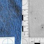
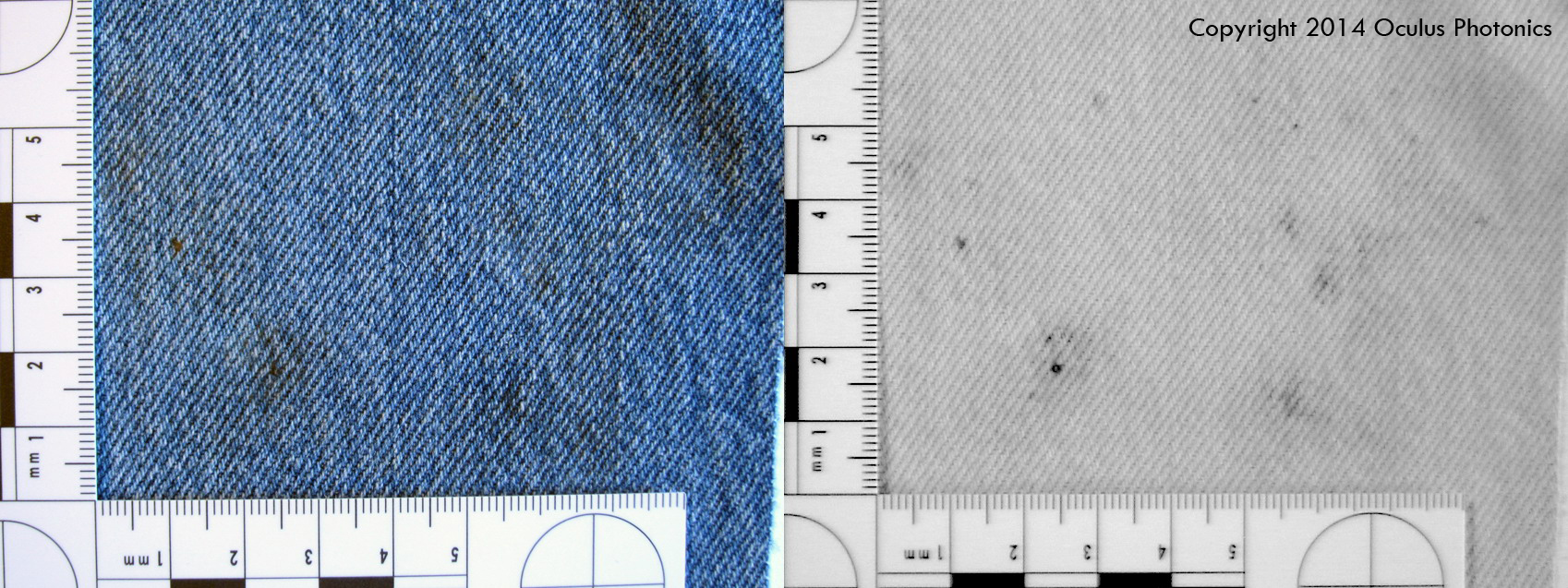
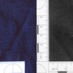
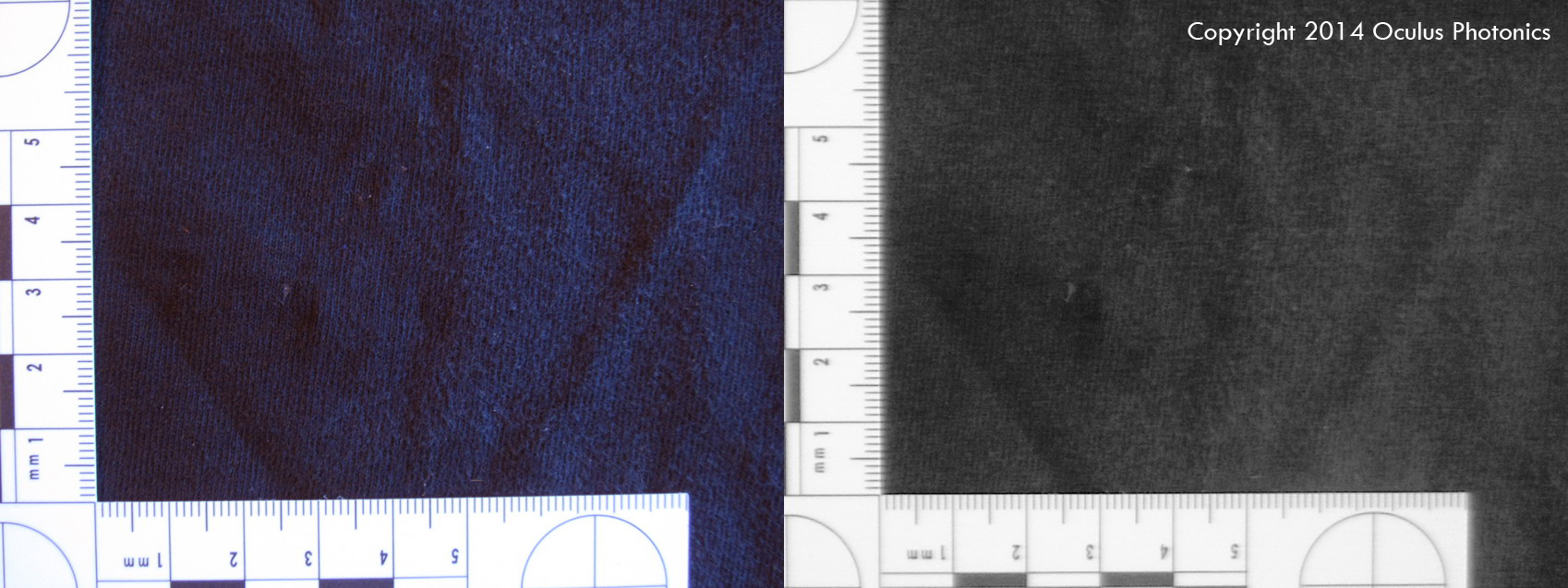
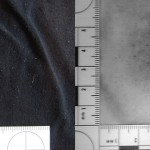
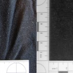
Just as with UV imaging, near-IR imaging sees crime scenes differently because near-IR light is absorbed and reflected differently than visible light. If you light up a crime scene with NIR rays, and image the NIR radiation that is reflected off the scene with a NIR camera, you will see things the eye can’t see. NIR radiation tends to penetrate through thin layers of materials, including the pigments and dyes used on textiles. For example, a black T-shirt made of cotton will look almost pure white to a NIR camera. The black dye molecules used on the cotton are essentially transparent to radiation around 850 nm in wavelengths. Any bloodstains or other residues on the T-shirt will often show up in a NIR image. A color photo or visual examination might show a faint indication of a bloodstain, but the evidence will be difficult to present in a convincing manner. A suspect wearing dark clothing that may have bloodstains can be rapidly examined with the SpectraScanner to find that evidence without the need for processing of the clothing.
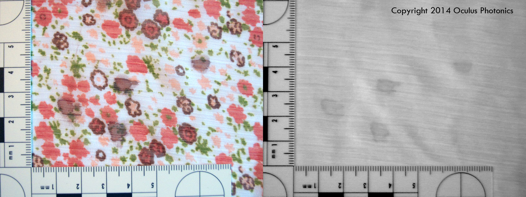
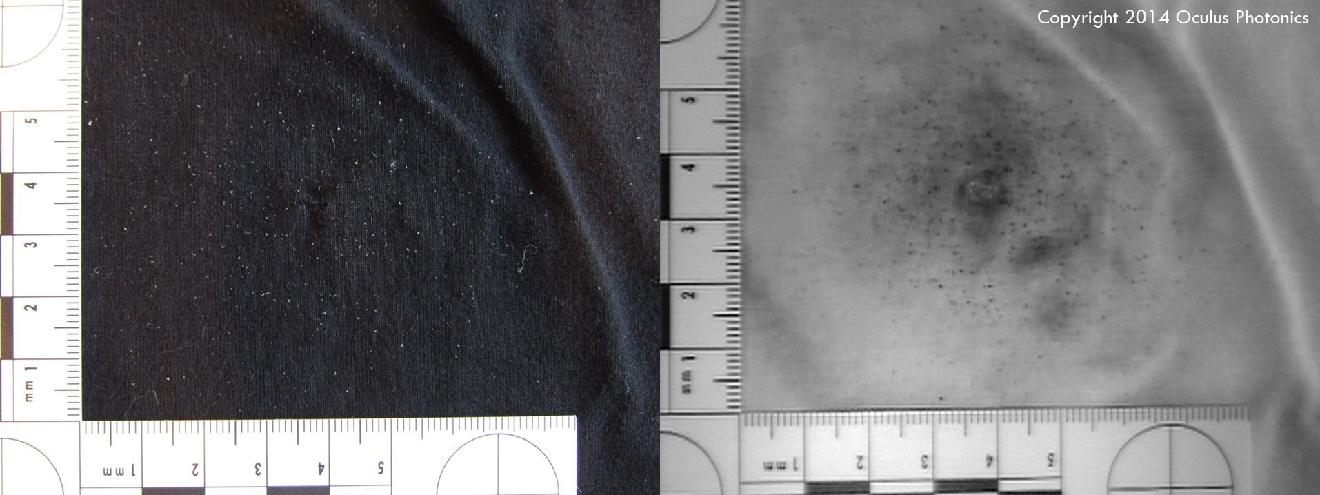
Gallery of bloodstain images on various textiles below (all samples courtesy of Rachel Leintz):
-
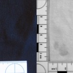
- Black cotton t-shirt
-
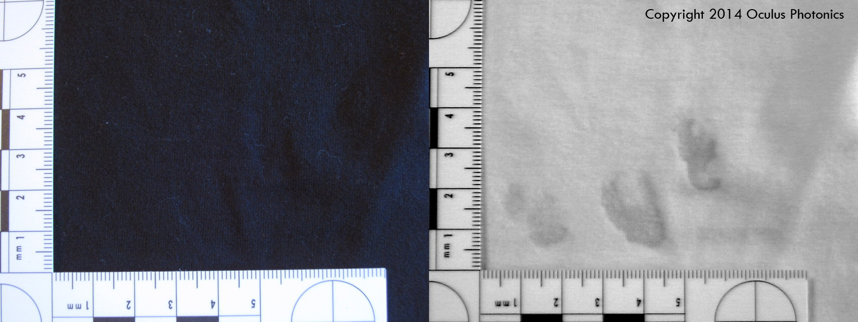
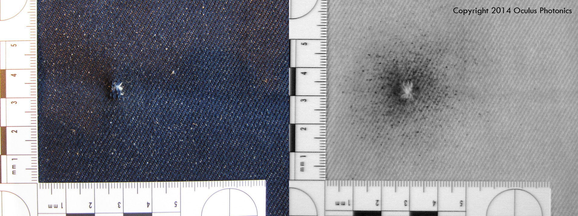
- Blue denim with bloody finger streaks
-
- Faded blue denim
-
- Black t-shirt. This fabric is dyed differently so that it does not look lighter in the NIR waveband, making it very hard to see the blood stains on it.
-
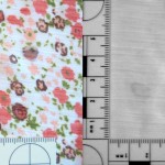
- Rayon blouse.
-
- Black cotton twill.
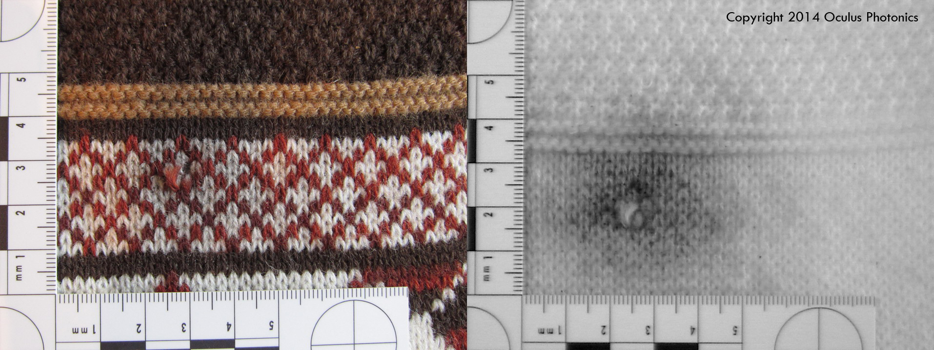
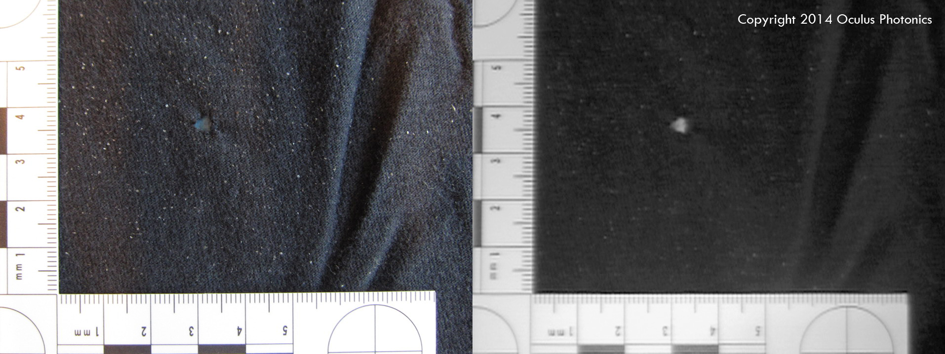
Gallery of muzzle blasts on textiles below:
-
- Black cotton twill.
-
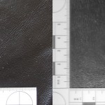
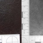
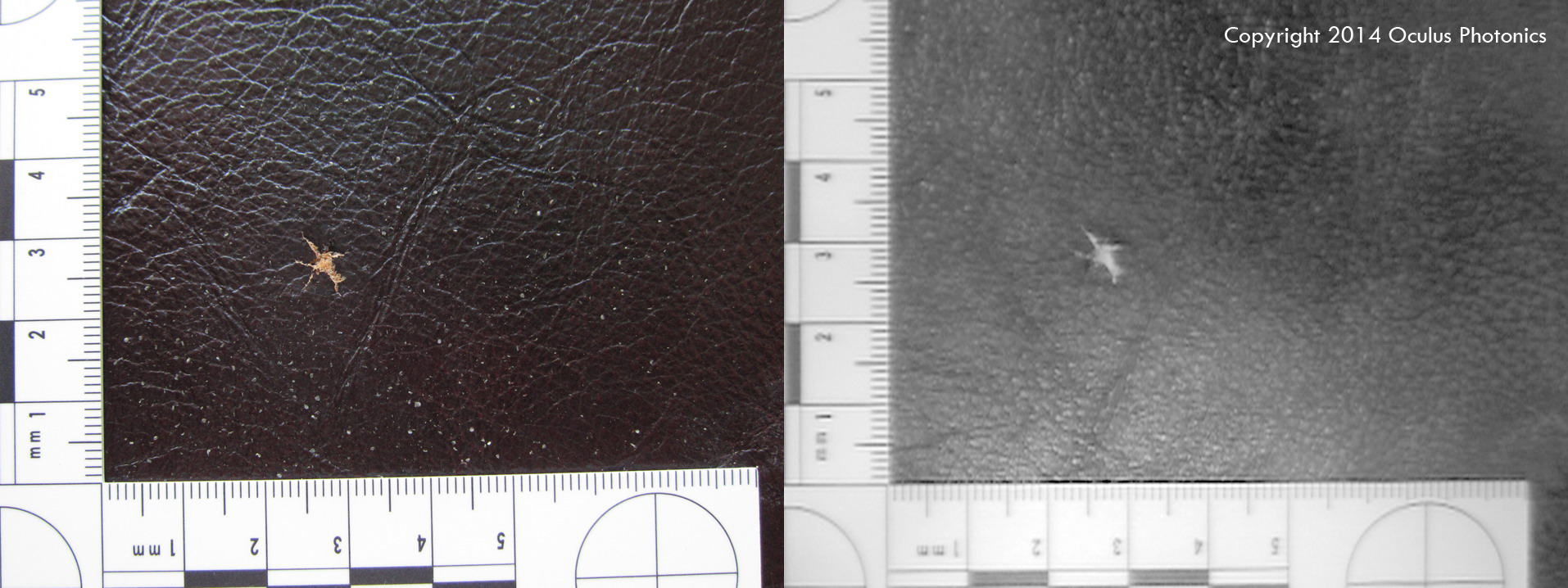
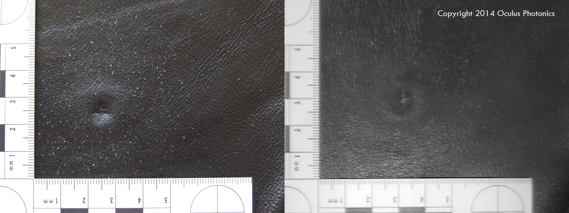
- Brown leather with shiny coating. Muzzle flash is not apparent because the surface does not lighten up in the NIR waveband.
-
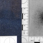
- Blue denim shot at close range.
-
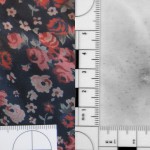
- Rayon blouse.
-
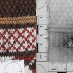
- Wool sweater.
-
- Blue denim.
-
- Black t-shirt.
-
- Black t-shirt. Fabric is colored with a pigment which does not lighten up in the NIR waveband.